17+ Plant Cell Microscope
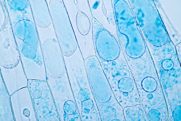
Two cells will be observed one from the skin of an onion and the other from a common aquarium water plant anacharis. Web Microscopy-based imaging approaches allow researchers to analyze the dynamic localization of cellular components membrane remodeling events the morphology and function of organelles the structural features of proteins and molecular complexes and their interaction networks.

Characterization Of Herpes Simplex Virus Type 1 L Particle Assembly And Egress In Hippocampal Neurones By Electron Cryo Tomography Ibiricu 2013 Cellular Microbiology Wiley Online Library
Web Through adaptation and application of super-resolution microscopy SRM methods sensitive enough to bring structural details at subcellular and supramolecular levels of both fixed and living cells astonishingly enhanced resolution of plant imaging can be achieved Komis et al 2018.

. Plants have three main organs. Web Comparing Plant Cells. Web Two types of light microscopes commonly are used in introductory plant pathology courses.
Plants cells differ from animal cells in that they have a cell wall which is glued to adjacent cells by the middle lamellae a large central vacuole and chloroplasts. The quality of cellular preservation is critical for both resolution and reliability of the imaging data. Web In this review we discuss sample preparation options for different 3D EM modalities and how their applications have transformed plant cell biology.
Web A typical animal cell is 1020 μm in diameter which is about one-fifth the size of the smallest particle visible to the naked eye. 18 June 2021 Biosensor imaging of a seedling measuring how the concentrations of the plant hormone gibberellin change as the plant grows. These leaf cells are commonly called liverworts with the scientific name.
Web Many cellular structures are too tiny to see by naked eyes. In this activity students section plant material and prepare specimens to view under a brightfield microscope. Compound microscopes and dissecting microscopes.
Roots stems and leaves. Examining specimens under a good microscope enables us to study these cellular structures and investigate their biological functions. A light microscope shines light on or through a specimen and uses a set of lenses to magnify and focus the image.
Web There are two general types of microscopes. Humans have been making use of plants for thousands of years. Web Methods to enhance plant cell outlines vary in complexity from straightforward imaging of cell wall autofluorescence to lengthy multistep processing for three-dimensional analysis of tissue architecture by confocal laser scanning microscopy CLSM eg 1 3.
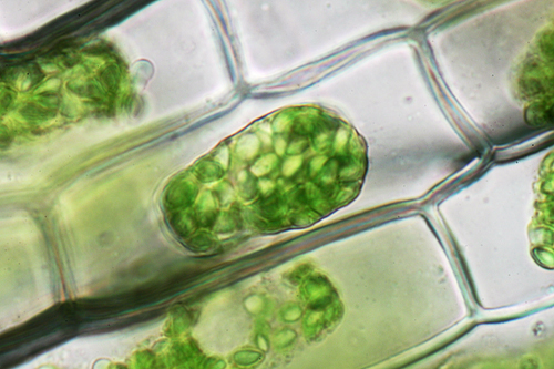
Review the principles of light microscopy and identify the major parts of the microscope. Take a look inside a plant leaf to see the chloroplasts and oil bodies hidden in this tiny world. In this article we will show you that you can study plant biology and anatomy using a premade slide set.
Web 69K views 3 years ago. Web As recently described by Marc Somssich in his short history of plant light microscopy the invention of the microscope and its use to observe plant tissues opened up a completely new world previously hidden to the human eye Somssich 2021. A new way to culture and image flowers is uncovering the processes that take place in reproductive cells buried deep in plants.
Students will compare both types of cells and identify structures visible in each. Sample preparation for 3D EM. Web We present a new large-scale three-fold annotated microscopy image dataset aiming to advance the plant cell biology research by exploring different cell microstructures including cell size.
Web 1 LAB 1. Most photographs of cells are taken using a microscope and these pictures can also be called micrographs. Web This protocol describes a detailed method for superresolution imaging of plant tissues by structured illumination microscopy SIM.
There are two general types of light microscopes. A diagram of a plant cell. Students will observe plant cells using a light microscope.
Web 12 Best Plant Cell Microscopes 2023 Top Models Reviewed. Web The microscope has been a powerful tool for studying cellular details and phenomena for hundreds of years ever since the first cells cork cells of plants were observed by Robert Hooke in the 1600s. January 3 2023 Leave a Comment.
Web A microscope is an instrument that magnifies objects otherwise too small to be seen producing an image in which the object appears larger. Light microscopes and electron microscopes. These microscopes are the compound microscope Figure 1 and the dissecting or stereo-microscope Figure 2.
Web In this review we focus on a selected set of microscopy techniques with powerful imaging capacity that have been successfully used to analyze growth and development in plants at multiple scales from the dynamics of proteins within cells to morphometric measurements of tissues. Spines hairs tendrils and thorns are usually modified. Introduction to the Microscope.
It was not until good light microscopes became available in the early part of the nineteenth century that all plant and animal tissues were discovered to be aggregates of individual cells. Microscopes Cells and Tissues Please bring your textbook to lab Objectives. First we introduce the basic principles behind.
Learn how to use the microscope to view slides of several different cell types including the use of the oil immersion lens to view bacterial cells. This lab will serve as an introduction to plant anatomy microscopy and the structure of a variety of plant tissues. Web Microscopy and stained specimens engage students visually as they learn about plant anatomy a topic covered in many biology and introductory science courses.
Easy to handle fun and interactive the best plant cell microscope will open up so many different avenues of biology for you. Dissecting microscopes are commonly used for the observation of larger objects and generally have magnifications of less than 100x. Details include microscope calibration tissue preparation.
Plant cell microscopes are a must-have if youre looking to build on your collection or want a microscope for the fun of it. Looking below the surface in plants.

Microscopic Photography Plant Cell Images Microscopic

278 Plant Cells Under Microscope Stock Photos High Res Pictures And Images Getty Images
3 577 Plant Cell Microscope Stock Photos Free Royalty Free Stock Photos From Dreamstime
Biology 130 Lab 3 Light Microscope Images
6 700 Plant Cell Microscope Stock Photos Pictures Royalty Free Images Istock Plant Cell Wall
Plant Cells Microscope Hi Res Stock Photography And Images Alamy

Plant Cells Under A Microscope Youtube

11 731 Plant Cell Microscope Images Stock Photos Vectors Shutterstock

Untitled Microscopic Photography Plant Cell Micro Photography

Beautiful Colored Micrographs Of Pollen Seeds And Plant Cells Moss And Fog
Biology 130 Lab 3 Light Microscope Images

6 700 Plant Cell Microscope Stock Photos Pictures Royalty Free Images Istock Plant Cell Wall

Cell 8 Pictures Of Plant Cells Under A Microscope Plant Cell Structure Under Microscope Plant And Animal Cells Plant Cell Structure Plant Cell
6 700 Plant Cell Microscope Stock Photos Pictures Royalty Free Images Istock Plant Cell Wall
Happy Plant Cells Under The Microscope R Pics

11 731 Plant Cell Microscope Images Stock Photos Vectors Shutterstock
6 700 Plant Cell Microscope Stock Photos Pictures Royalty Free Images Istock Plant Cell Wall